Leave message
Can’t find what you’re looking for?
Fill out this form to inquire about our custom protein services!
Inquire about our Custom Services >>


 Order Online! Now! Get your $50 coupon for online order.
 Order Online! Now! Get your $50 coupon for online order.
Request a FREE sample of our GMP products! Request a FREE sample of our GMP products!
Fill out organ-on-a-chip questionnaire to win a FREE gift! Fill out organ-on-a-chip questionnaire to win a FREE gift!
> Lymphocyte-activation gene 3 (LAG-3) The clinical success of several anti-PD1 and anti-CTLA4 antibodies has boosted the research of other immune checkpoint proteins. The lymphocyte-activation gene 3 (LAG-3), is of particular interest to researchers because it functions differently from the canonical negative checkpoint regulators such as PD1 and CTLA4.
LAG-3 is expressed on cell membranes of natural killer cells (NK), B cells, tumor-infiltrating lymphocytes (TIL), T cells, and dendritic cells (DC), and it appears to play complicated roles in the immune pathway. The binding between soluble LAG-3 (sLAG-3) and MHCII molecules promotes the maturation of dendritic cells, while the interaction between MHCII and LAG-3 on membrane leads to the negative regulation of T-cell activity.
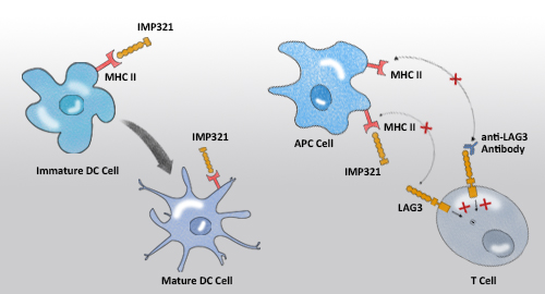
Several therapeutic agents have been developed to target the LAG-3 pathway. IMP321, a sLAG-3-Fc fusion protein developed by Prima Biomed, is one of the leading drugs that currently being investigated in a phase II clinical trial. IMP321 augments immune response against cancer cells through two distinct mechanisms. It binds with MHCII molecules on the immature dendritic cells to enhance tumor antigen presentation. At the same time, it also blocks the MHCII molecule to bind with LAG-3-expressing CD8+ T-cells, and by so doing prevents the T-cells from being negatively regulated. Other investigative drugs, such as Bristol-Myers Squibb's anti-LAG-3 antibody BMS-986016, directly bind with LAG-3 expressed on the T-cells and block the LAG-3-MHCII interaction. As a result, the T-cells can be activated to boost immune response.
ACROBiosystems provides a comprehensive panel of LAG-3 proteins, including Cynomolgus LAG-3, Mouse IgG2a Fc Tag (Cat. No. LA3-C52A0), Cynomolgus LAG-3, Fc Tag (Cat. No. LA3-C5252), and Human LAG-3, Fc Tag (Cat. No. LA3-H5255).
| Cat. No. | Species | Product Description | Structure | Purity | Features |
|---|---|---|---|---|---|
| LA3-H5222 | Human LAG-3 / CD223 Protein, His Tag |  | |||
| LA3-H52Aa | Human LAG-3 / CD223 Protein, Mouse IgG2a Fc Tag |  | |||
| LA3-H5255 | Human LAG3 / CD223 Protein, Fc Tag (HPLC-verified) |  | |||
| LA3-H82Fb | Biotinylated Human LAG-3 / CD223 Protein, Fc,Avitag™ |  | |||
| LA3-M52H5 | Mouse LAG-3 / CD223 Protein, His Tag |  | |||
| LA3-H82F3 | Biotinylated Human LAG-3 / CD223 Protein, Mouse IgG2a Fc,Avitag™ |  | |||
| LA3-C5252 | Cynomolgus LAG-3 / CD223 Protein, Fc Tag |  | |||
| LA3-C52A0 | Cynomolgus LAG-3 / CD223 Protein, Mouse IgG2a Fc Tag |  | |||
| LA3-H525c | Human LAG-3 / CD223 Protein, Llama IgG2b Fc Tag, low endotoxin |  | |||
| LA3-H522a | Human LAG-3 / CD223 Protein, His Tag, low endotoxin |  | |||
| LA3-R52H5 | Rat LAG3 / CD223 Protein, His Tag |  | |||
| LA3-M52Ha | Marmoset LAG-3 / CD223 Protein, His Tag |  |
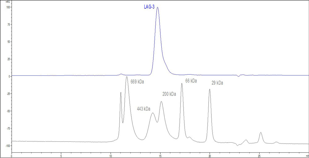
Fig.1 The purity of Human CD73, His Tag (HPLC-verified) (Cat. No. CD3-H52H7) was greater than 90% as determined by SEC-HPLC.
FACS analysis of LAG-3 mFc binding to Daudi cell surface
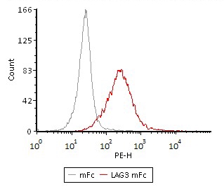
Fig.2 Flow Cytometry assay shows that Human LAG3, Mouse IgG2a Fc Tag (Cat. No. LA3-H52Aa) can bind to Daudi cell surface. The concentration of LAG3 used is 0.3 μg/mL.
Competitive experiment of neutralizing anti-LAG-3 antibody
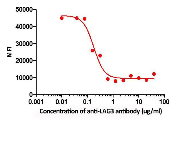
Fig.3 FACS analysis shows that the binding Human LAG3, Mouse IgG2a Fc Tag (Cat. No. LA3-H52Aa) to Daudi cells was inhibited by increasing concentration of neutralizing Anti-LAG3 antibody. The concentration of LAG3 used is 1 μg/mL. The IC50 is 0.165 μg/mL.
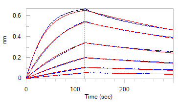
Fig.4 Loaded Anti-LAG-3 mAb (Human IgG1) on AHC Biosensor, can bind Human LAG-3, His Tag (Cat. No. LA3-H5222) with an affinity constant of 7.47 nM as determined in BLI assay (ForteBio Octet Red96e).
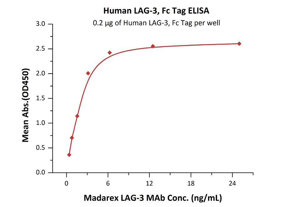
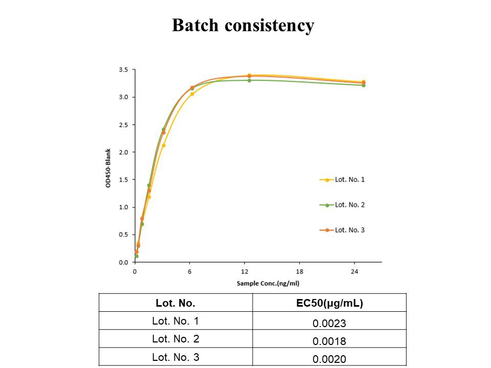
Fig.5 Immobilized Human LAG-3, Fc Tag (HPLC-verified) (Cat. No. LA3-H5255) at 2 μg/mL (100 μL/well) can bind Madarex LAG-3 mAb with a linear range of 0.4-3 ng/mL. The ELISA date showed that the Human LAG-3 protein has high batch-to-batch consistency.

This web search service is supported by Google Inc.


 A-Z
A-Z