


 Sep 17, 2026
 Page Views:127


Introduction
In ADC development and antibody functional research, internalization efficiency and intracellular trafficking are core developability attributes. Labeling requirements also differ from one experimental stage to the next: high-throughput sample screening prioritizes ease of operation, while studies involving antibodies from unconventional species or rare subclasses call for broader compatibility and greater labeling stability.
To support these different internalization assay scenarios, ACROBiosystems has launched the pHintra™ Antibody Labeling Kit. The kit uses an amine-based covalent labeling strategy to efficiently couple a pH-sensitive fluorescent dye to IgG antibodies from multiple species and subclasses. After a simple spin-column purification, the labeled antibody can be used directly in antibody internalization assays based on flow cytometry or cell imaging — a more flexible detection tool for antibody and ADC research.
How It Works
The pHintra™ Antibody Labeling Kit provides a rapid, simple method for covalently attaching a pH-sensitive dye to antibodies through primary amine groups on the antibody surface. The labeled antibody can then be used to measure antibody internalization efficiency by flow cytometry or cell imaging.
The pH-sensitive dye (Em660) emits bright fluorescence in acidic environments while keeping background noise low, with an excitation wavelength of 643 nm and an emission wavelength of 660 nm.
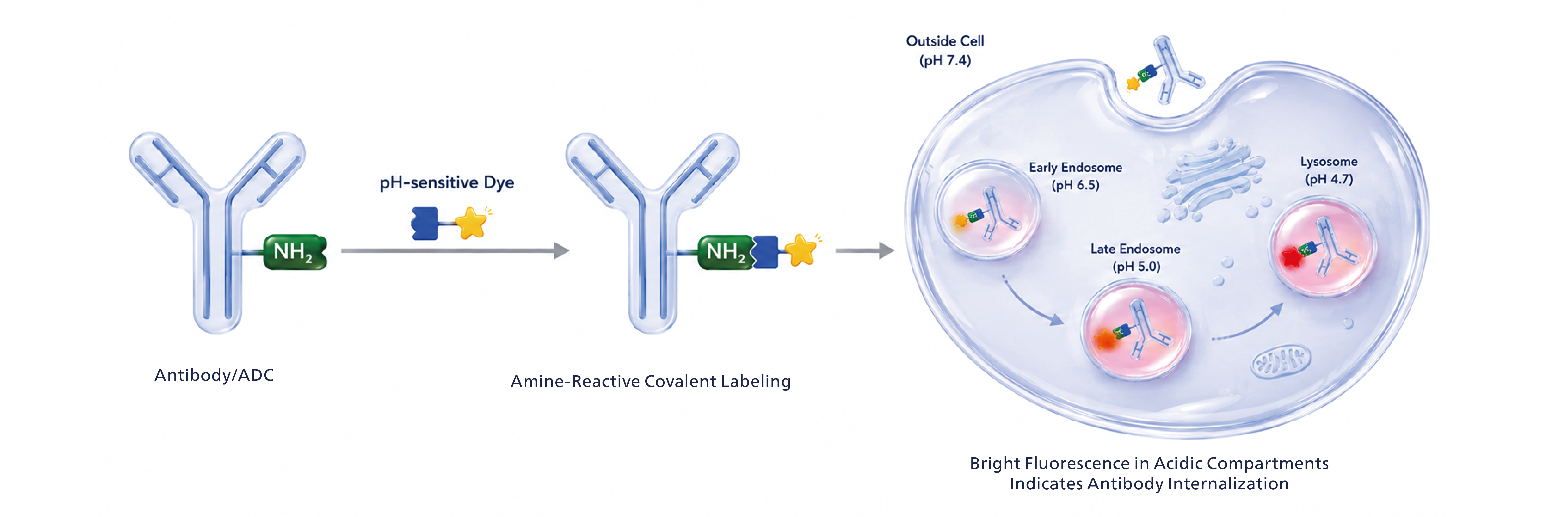
Figure: Technical principles of pHintra™ Antibody Labeling Kit
The dye is minimally fluorescent at neutral pH. Outside the cell (pH 7.4), a labeled antibody binds its antigen on the cell surface. Once the antibody is internalized by endocytosis, it is routed from early endosomes (pH 6.5) through late endosomes (pH 5.0) to lysosomes (pH 4.7). As the vesicle lumen gradually acidifies, the dye is activated and emits bright fluorescence — providing a direct readout that the antibody has completed cellular internalization.
Product Information
|
Cat. No. |
Product Description |
Order |
|---|---|---|
|
pH-Sensitive Fluorescent Dye Antibody Labeling Kit (Em660, 100 μg/Reaction) |
Key Features
- Direct labeling, no secondary antibody. The pH-sensitive dye is covalently attached to the target antibody, adding only a small increase in molecular weight and improving assay flexibility.
- High signal-to-noise ratio. Strong fluorescence with low background in acidic environments improves the sensitivity of internalization detection.
- Acid-responsive fluorescence. The dye brightens only in acidic intracellular compartments such as endosomes and lysosomes, faithfully reporting true antibody internalization.
- Just 30 minutes of hands-on time. Antibody labeling requires only about 30 minutes of manual work, and the full experiment is completed in about 2.5 hours.
- Broad antibody compatibility. Works with antibodies from all species and across subclasses, fitting more experimental scenarios.
- All-in-one reagent set. All required reagents are provided in a single kit with a standardized workflow, eliminating method development.
Labeling Workflow at a Glance
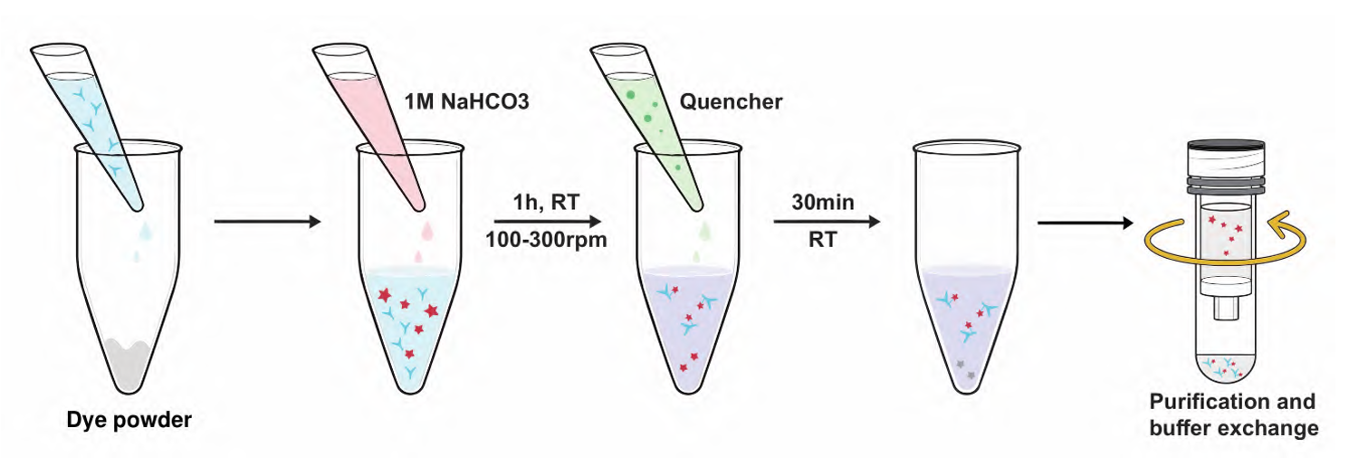
Figure: Quick guide — antibody labeling workflow
Download the datasheet (DS) for the detailed protocol
Validation Data
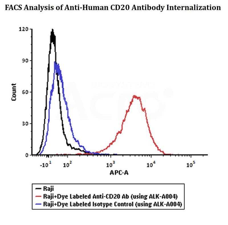
Labeled Antibodies Perform in FACS Internalization Assays
|
|
|
|---|---|
|
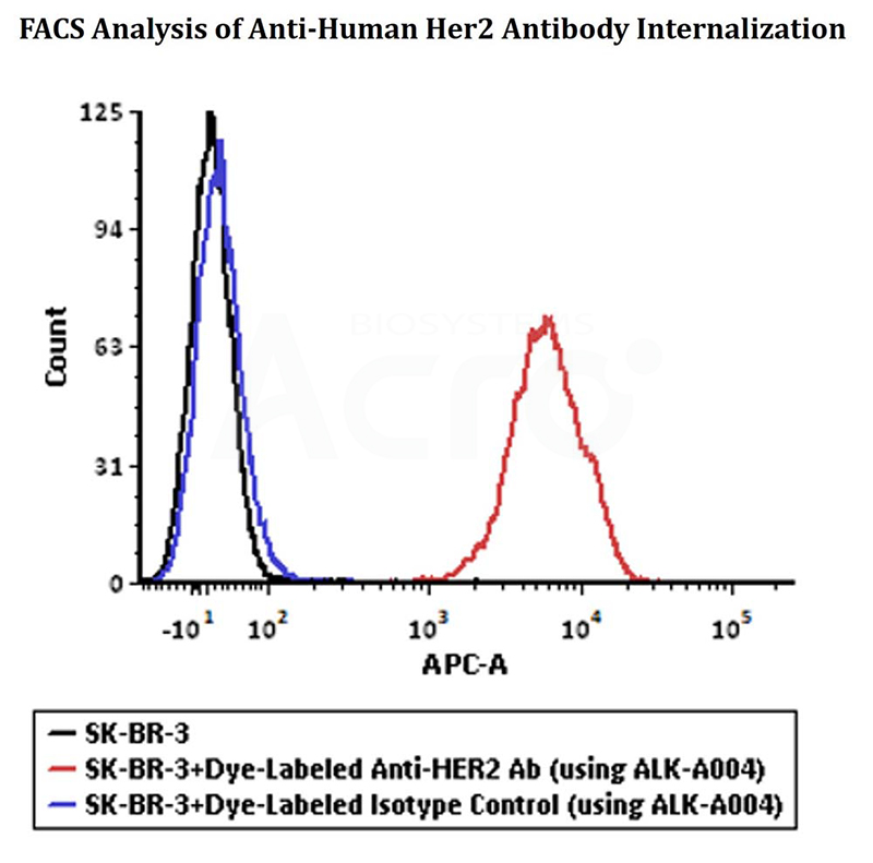
Anti-Her2 Ab and Human IgG1 isotype control were labeled using pH-Sensitive Fluorescent Dye Antibody Labeling Kit (Cat. No. ALK-A004). SK-BR-3 cells were treated with Dye Labeled Anti-Her2 Ab and Dye Labeled Isotype Control separately at 37℃ for 4 hours, then analysis by Flow cytometric. APC signal was used to evaluate the internalization signal. |
Anti-CD20 Ab and Human IgG1 isotype control were labeled using pH-Sensitive Fluorescent Dye Antibody Labeling Kit (Cat. No. ALK-A004). Raji cells were treated with Dye Labeled Anti-CD20 Ab and Dye Labeled Isotype Control separately at 37℃ for 4 hours, then analysis by Flow cytometric. APC signal was used to evaluate the internalization signal. |
Labeled Antibodies Perform in Fluorescence Imaging Internalization Assays
Application of pH-Sensitive Fluorescent Dye Antibody Labeling Kit in Endocytosis Detection
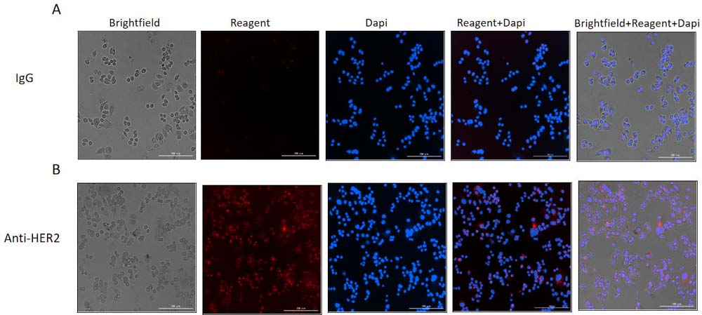
SK-BR-3 cells were treated with Anti-Her2 Abs-pH-Sensitive Fluorescent Dye Antibody Labeling Kit conjugate and IgG1 Isotype-pH-Sensitive Fluorescent Dye Antibody Labeling Kit conjugate separately for 20 hours (red), then stained with NucBlue Live ReadyProbes (blue) for 20 minutes and imaged on the Cytation 5. A. IgG1 Isotype-pH-Sensitive Fluorescent Dye Antibody Labeling Kit conjugate. B. Anti-Her2 Abs-pH-Sensitive Fluorescent Dye Antibody Labeling Kit conjugate.
Related Products
Working with a human IgG antibody? Try the ready-to-use internalization detection reagent.
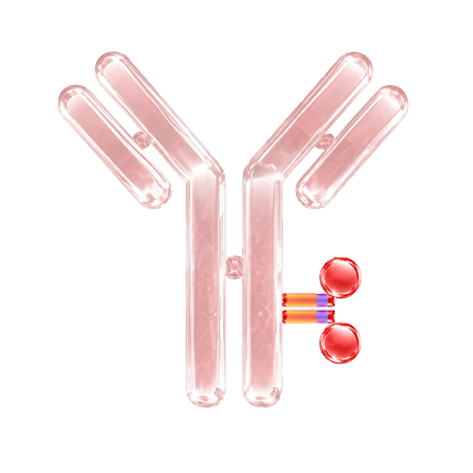
pHintra™ Antibody Internalization Detection Reagent (Cat. No. IGG-PZF2001), featuring a pH-sensitive red fluorescent dye that labels the human IgG Fc region. The reagent forms a stable fluorescent complex within 10 minutes, enabling rapid assessment of antibody internalization processes. View product details →
Choose the Internalization Assay Format That Fits
| Category |
 Ready-to-Use Internalization Assay Ready-to-Use Internalization Assay
|
 Labeling Internalization Assay Labeling Internalization Assay
|
|---|---|---|
 Labeling Method Labeling Method
|
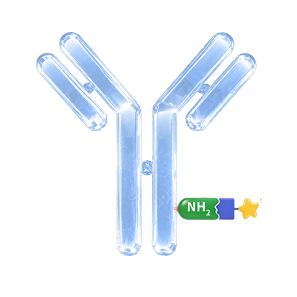
Fc-specific non-covalent labeling | Primary amine-based covalent labeling |
 Antibody Compatibility Antibody Compatibility
|
Human IgG antibodies & ADCs | Antibodies/ADCs from different species and subclasses |
 Preparation Time Preparation Time
|
Approximately 10 min antibody labeling | 30 min hands-on work |
 Detection Method Detection Method
|
Flow cytometry / Cell imaging | Flow cytometry / Cell imaging |
 Best Suited For Best Suited For
|
|
|
 Core Advantage Core Advantage
|
No purification required, approximately 10 min labeling | Broad compatibility across antibody species and subclasses, as well as ADCs |
Not sure which format fits your workflow? [Contact us →]
Explore Solutions for ADCs Development