Leave message
Can’t find what you’re looking for?
Fill out this form to inquire about our custom protein services!
Inquire about our Custom Services >>


> Insights > S protein is in a trimeric form as verified by three methods from NIH SARS-CoV-2 is a positive-sense single-stranded RNA virus that is very contagious in humans. The viral envelope consists of a lipid bilayer, in which the membrane (M), envelope (E) and spike (S) structural proteins are anchored. The spike protein is a homotrimer, which is composed of an S1 and S2 subunit. The globular S1 subunit is involved in receptor recognition, whereas the S2 subunit facilitates membrane fusion and anchors S into the viral membrane. Therefore, the S protein is the key protein that the coronavirus uses to invade human cells. S protein is also the main antigen that causes the host immune system to produce neutralizing antibodies after infection.
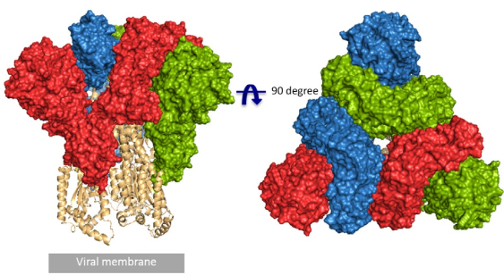
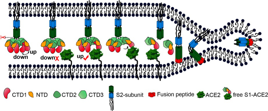
According to the article published in Science earlier this year, a team from UT Austin determined the cryo-electron microscopy structure of the 2019-nCoV S trimer in the prefusion conformation. [1] As shown in Fig.1, prefusion S glycoproteins adopt a similar mushroom-like homo-trimer architecture, of which the stem is mainly composed of three S2 subunits, and the top cap consists of three interwoven S1 subunits.[3] Another team from Tsinghua University confirmed that the conformational switch of the receptor-binding domain(RBD) from the “down” to “up” position is a prerequisite for receptor binding (Fig. 2). Therefore, the homo-trimer architecture is essential to achieve the complete function of the S protein.

Fig. 1 Front and top view of the trimeric coronavirus spike protein ectodomain obtained by cryo-electron microscopy analysis. Three S1 protomers (surface presentation) are colored in red, blue, and green. The S2 trimer (cartoon presentation) is colored in light orange. (Source: R.J.G. Hulswit, et al., Coronavirus Spike Protein and Tropism Changes. Adv Virus Res. 2016; 96: 29–57.)


Fig. 2 S protein mediates viral membrane and membrane fusion by binding to host cell receptor ACE2. (Source: Song W, Gui M, Wang X, Xiang Y (2018) Cryo-EM structure of the SARS coronavirus spike glycoprotein in complex with its host cell receptor ACE2. PLoS Pathog 14(8): e1007236.)
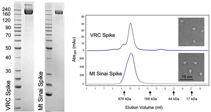
The CoV spike (S) glycoprotein is a key target for vaccines, therapeutic antibodies, and diagnostics. The correct trimeric structure and functionality of the recombinant S protein reagent are important. Therefore, strict quality control is required in terms of the molecular weight and aggregation state. In a recent work from NIH, the scientist used three ways including SDS-PAGE, analytical size exclusion chromatography, and transmission electron microscopy to verify that the recombinant S protein is in a trimeric form when developing a COVID-19 serological diagnostic kit (Fig. 3).

Fig. 3 Verification of Spike trimer structure using SDS-PAGE, analytical size exclusion chromatography and transmission electron microscopy
(Source: Carleen Klumpp-Thomas, et al. Standardization of enzyme-linked immunosorbent assays for serosurveys of the SARS-CoV-2 pandemic using clinical and at-home blood sampling, preprint at https://www.medrxiv.org/content/10.1101/2020.05.21.20109280v1.)
ACROBiosystems has specially designed a highly active S trimer protein. This is the only verified trimeric S protein on the market. Our SEC-MALS and negative-stain EM data reveal that ACRO's S protein is in the correct trimer form under physiological conditions, and purity is over 80%. The Biacore affinity test shows that the S trimer protein has an affinity constant as 35.3nM to human ACE-2 protein, which is similar to the value in the reference.[1]
>>> SPR

Fig 5. Human ACE2, Fc Tag (Cat. No. AC2-H5257) Captured on CM5 chip via anti-human IgG Fc antibodies surface can bind SARS-CoV-2 (COVID-19) Full Length S protein (R683A, R685A), His Tag, active trimer (Cat. No. SPN-C52H8) with an affinity constant of 35.3 nM as determined in a SPR assay (Biacore 8K).
Protocol
Reference:
1. Wrapp D, et al., Cryo-EM structure of the 2019-nCoV spike prefusion conformation. Science, 2020 Mar 13;367(6483):1260-1263.
2. R.J.G. Hulswit, et al., Coronavirus Spike Protein and Tropism Changes. Adv Virus Res. 2016; 96: 29–57.
3. Song W, Gui M, Wang X, Xiang Y (2018) Cryo-EM structure of the SARS coronavirus spike glycoprotein in complex with its host cell receptor ACE2. PLoS Pathog 14(8): e1007236.
4. Carleen Klumpp-Thomas, et al. Standardization of enzyme-linked immunosorbent assays for serosurveys of the SARS-CoV-2 pandemic using clinical and at-home blood sampling, preprint at https://www.medrxiv.org/content/10.1101/2020.05.21.20109280v1.
This web search service is supported by Google Inc.


