Quantitative Analysis of mAb in Serum
 Publication Date:2018-04-18
 Page Views:46125
Introduction
Pharmacokinetics (PK) is a branch of science dedicated to the quantitative analysis of absorption, distribution, metabolism and excretion of drug molecules within the body of a living organism. All pre-clinical and clinical studies include the measurement of serum drug concentration, both in animals and in patients, at different points after drug administration. The result is an important indicator of the drug’s pharmacokinetic properties and is pertinently relevant to dosing recommendations.
Case Study
The booming market of biologic drugs, driven by a flurry of success with monoclonal antibodies, brings the need for standard high throughput assays to evaluate the content of mAbs in serum samples. There are several types of assay designs based on different principles. The table below listed three most common ELISA setups for serum antibody detection, and their general advantages and disadvantages. To evaluate their performance in real applications, we tested them in the following case studies measuring the concentration of anti-PD1 mAb in serum samples.
| Method | Coating | Sample | Secondary Antibody | Advantage | Disadvantage | Case Studies |
|---|---|---|---|---|---|---|
| Indirect assay | Antigen | Serum | Goat anti-human IgG | Simple | High background | Case I |
| Antibody-directed competitive assay | Antigen | Serum with labeled competition antibody | SA-HRP | Low background | Additional reagent/labeling required | Case II |
| Ligand-directed competitive assay | Natural ligand for the antigen | Serum with labeled PD-1 | SA-HRP | Low background | Narrow detection range | Case III |
Please feel free to contact us by pk@acrobiosystems.com if you have any questions.
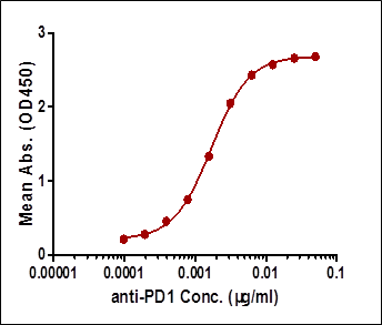
Case I: Indirect ELISA
-
Equipment:BMG CLARIOstar microplate reader
-
Sample:Serum samples containing a monoclonal PD-1 antibody
-
Main Reagents:
Recombinant Human PD-1 protein (Cat. No. PD1-H5221 , ACROBiosystems, Newark, DE, USA)Peroxidase AffiniPure Goat Anti-Human IgG, Fcγ Fragment Specific (Cat. No. 109-035-098, Jackson Lab, Bar Harbor, ME, USA) -
Protocol:
1. Coat the microplate with 0.1 µg/well rhPD-1 for 16hr;2. Prepare serial sample dilutions (1:2);3. Wash the plate;4. Add samples 100 µl onto the plate;5. Wash the plate;6. Add HRP-conjugated Goat Anti-Human IgG 0.05 µg/ml;7. Add TMB for colorimetric detection. -
Protocol:
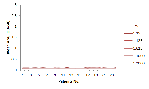
To avoid unspecific binding, all serum samples was diluted by a factor of 1000 before assays. The detection range is 0.19-6.25 µg/mL, and the sensitivity is 0.19 µg/mL (Figure 1).

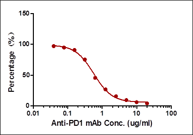
Case II: Antibody-directed competitive ELISA
-
Equipment:BMG CLARIOstar microplate reader
-
Sample:Serum samples containing a monoclonal PD-1 antibody
-
Main Reagents:
ELISA Assay Kit for Anti-PD-1 h-mAb in Human Serum (Cat. No. EPH-V1, ACROBiosystems, Newark, DE, USA) -
Protocol:
1. Coat the microplate with 0.1 µg/well rhPD-1 for 16hr;2. Prepare serial sample dilutions (1:2);3. Mix samples with the biotinylated PD-1 antibody provided in the kit to a final concentration of 10%;4. After washing, add the mixed samples 100 µl from step 2 to the wells;5. After washing, add HRP conjugated Streptavidin 0.1 µg/ml;6. Add TMB for colorimetric detection. -
Result:
The detection range is 0.78-25 µg/mL and the sensitivity are 0.78 µg/mL (Figure 2). -
Related Products:
ELISA Assay Kit for Anti-PD-1 h-mAb in Human Serum (Cat. No. EPH-V1 )

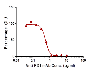
Case III: Ligand-directed competitive ELISA
-
Equipment:BMG CLARIOstar microplate reader
-
Sample:Serum samples containing a monoclonal PD-1 antibody
-
Main Reagents:
Recombinant human PD-L1-Fc protein (Cat. No. PD1-H5258, ACROBiosystems, Newark, DE, USA)Biotinylated human PD-1 (Cat. No. PD1-H82F2, ACROBiosystems, Newark, DE, USA)HRP-conjugated Streptavidin Protein (Cat. No. 21126, Thermo Fisher Scientific) -
Simplified Protocol:
1. Coat the microplate with 0.2 µg/well rhPD-L1 for 16hr;2. Prepare serial sample dilutions (1:2), and mix with biotinylated PD-1 to a final concentration of 10%;3. After washing, add the samples 100 µl from step 2 to the wells;4. After washing, add HRP conjugated Streptavidin 0.1 µg/ml;5. Add TMB for colorimetric detection. -
Result:
The detection range is 1.565-6.25 µg/mL and the sensitivity are 1.565 µg/mL (Figure 3).

Methods Comparison
| Case | Detection Range (µg/mL) | Sensitivity (µg/mL) | Specificity |
|---|---|---|---|
| Case I:Indirect ELISA | 0.19-6.25 | 0.19 | High background |
| Case II:Antibody-directed competitive ELISA | 0.78-25 | 0.78 | Very low background |
| Case III:Ligand-directed competitive ELISA | 1.565-6.25 | 1.565 | Very low background |
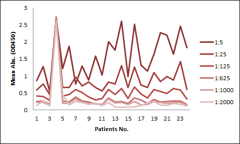
Among the aforementioned three assay designs, the ligand-directed competitive ELISA (Case III) has the narrowest detection range and lowest sensitivity, indicating that it’s not the best solution for the application. The sensitivity for the indirect ELISA is the best. However, the use of goat anti-human IgG as secondary antibody results in high background due to unspecific binding, and therefore require pre-dilution before analyses (Fig. 4A). On the other hand, the antibody-directed competitive ELISA uses HRP-conjugated Streptavidin for secondary detection, which minimize the background interference (Fig. 4B). In addition, it also has the widest detection range among the group, although the sensitivity is lower than the indirect method.
In most applications, the background issue is a bigger concern than detection sensitivity, as a detection sensitivity of less than 1 µg/ml is already good enough. Therefore, we opt to use the antibody-directed competitive ELISA for the PK kits.
We have launched kits for the studies of CTLA-4, PD-1, and HER-2, respectively, in both humans, and common experimental animals.
For more information
 Popular ArticlesRelated RecommendationsPopular Events
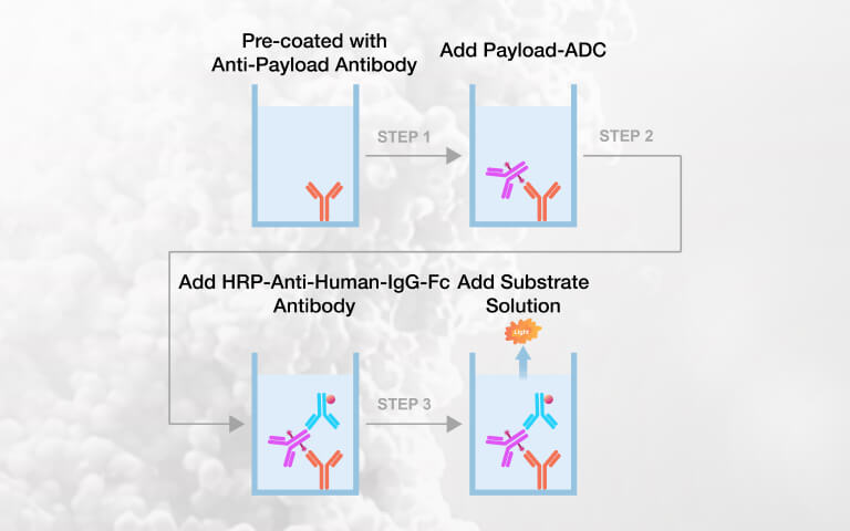 Payload-Specific ELISA Kits for Intact ADC Quantification in Preclinical PK Studies2026-07-22Page Views:57
 Expanding the Potential of Hematopoietic Stem Cells (HSCs): Emerging Strategies for HSCs Expansion and Clinical Manufacturing2026-07-22Page Views:42
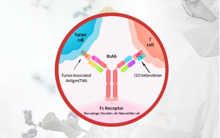 Advances and Challenges of BCMA×CD3 Bispecific Antibodies in Multiple Myeloma2026-07-21Page Views:106
 E. coli Host Cell DNA Testing: Independent Eurofins Validation Demonstrates Reliable Residual DNA Quantification2026-07-20Page Views:90
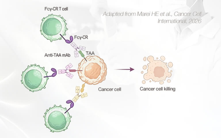 What Determines FcγR-CR T Function Beyond the Therapeutic Antibody?2026-07-14Page Views:65


![[Free Sample] Universal Fluorescent Anti-VHH Antibody — Precise Detection of VHH-Based CAR Expression](https://console.acrobiosystems.com/uploads/demo/2026/06/02/Anti-VHHAntibodySmallbanner.jpg)
 Related Products