Evaluation of CAR Expression
 Publication Date:2021-05-24
 Page Views:54589

Evaluating CAR expression is an essential step in the production of CAR-T cells. This is often done by flow cytometry,using protein L, anti-Fab antibodies, anti-idiotypic antibody or target antigens as detection antibodies. Among these common choices, target antigens are widely considered to be the best option, because it offers high specificity and minimal background staining.
Click to download the Guidance for CAR detection
| Reagents | Mechanism | Pros | Cons |
|---|---|---|---|
| Target Antigens |
|
|
|
| Protein L |
|
|
|
| Anti-Fab antibody |
|
|
|
| Anti-idiotypic Antibody |
|
|
|
Please feel free to contact us by cart@acrobiosystems.com if you have any questions.
Case Studies
Case No.1 Evaluation of anti-BCMA CAR expression using biotinylated BCMA
-
Reagents:Biotinylated human BCMA protein, Fc & Avi Tag (ACROBiosystems, Cat. No. BC7-H82F0); PE Streptavidin (Biolegend, Cat. No. 405204).
-
Samples:Jurkat cells expressing BCMA-CAR ; Primary T cells expressing BCMA-CAR
-
Brief Protocol:
1.Transduce human primary T cells or Jurkat T cells with BCMA-CAR lentivirus.2.Collect the CAR transduced human T cells and un-transduced T cells.3.Count the cells and aliquot 2 x10⁵ cells for staining into each tube.4.Wash the cells once with a FACS buffer.5.Dilute the biotinylated human BCMA protein (ACROBiosystems, Cat. No. BC7-H82F0) with working stock to 3µg/ml. Add 100µl of Biotinylated BCMA to each tube and incubate for 60 minutes in ice (protect from light).6.Wash the cells with FACS buffer once and stain with 100µl of 1µg/ml PE conjugated streptavidin for 30 minutes in ice (protect from light).7.Wash the cells twice with FACS buffer.8.Resuspend the cells in 200µl FACS buffer.9.Analyze the results using Attune NxT flow cytometer and FlowJo software. -
Results:The data show that BCMA-CAR expression can be successfully quantified using Biotinylated BCMA from ACROBiosystems (Cat. No. BC7-H82F0) both in jurkat cell line (Fig.1) and primary T cells (Fig.2). Control cells lacking BCMA-CAR expression showed no positive staining (Fig. 3) with PE- Streptavidin conjugate indicating that the staining using biotinylated BCMA is specific.
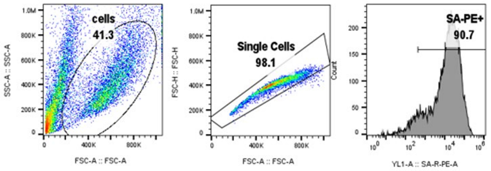
Fig. 1. BCMA-CAR expression in Jurkat cells
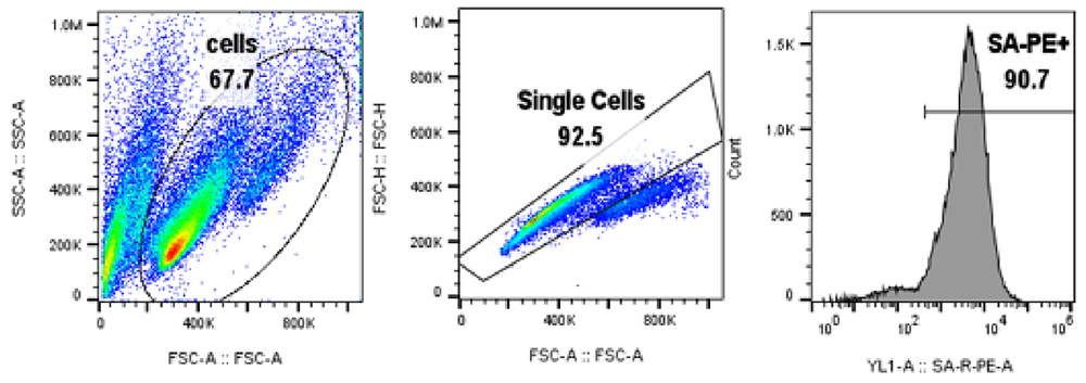
Fig. 2. BCMA-CAR expression in Primary T cells
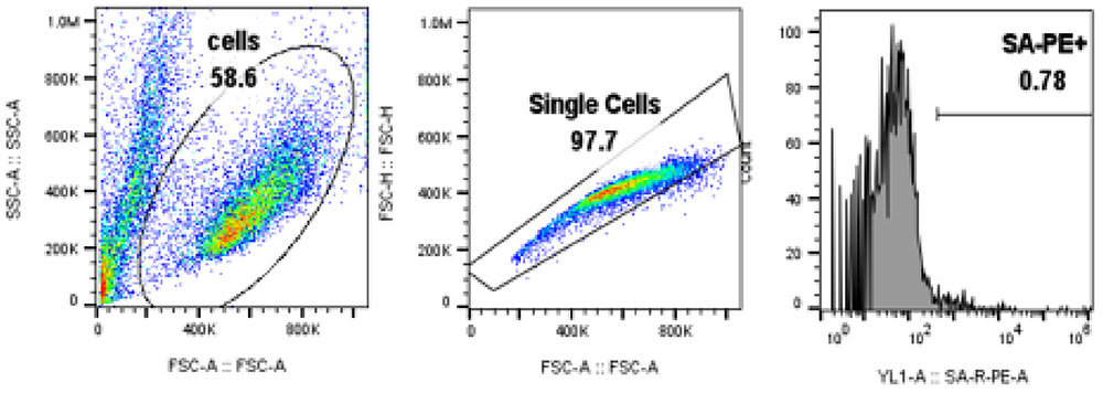
Fig. 3 Control cells without BCMA-CAR expression.
Data are kindly provided by R&D team, Theragent Inc 
-
Reagents:Biotinylated human BCMA protein, Fc & Avi Tag (ACROBiosystems, Cat. No. BC7-H82F0);PE Streptavidin (Biolegend, Cat. No. 405204).
-
Samples:Anti-BCMA CAR-transduced human primary T-cells.
-
Brief Protocol:
1.Transduce human primary T-cells with a lentiviral vector to express anti-BCMA CAR;2.Three days post-transduction, cells were stained with a biotinylated human BCMA protein (ACROBiosystems, Cat. No. BC7-H82F0);3.After washing, secondary labeling was performed with PE Streptavidin;4.The cells were analyzed using BD FACSCaliburTM flow cytometer, and the data was analyzed with FCS Express 6 Plus software. -
Results:The data showed that the expression of anti-BCMA CARs on transduced T cell surface from donor 1 and donor 2 were 52.72% and 73.49%, respectively.
-
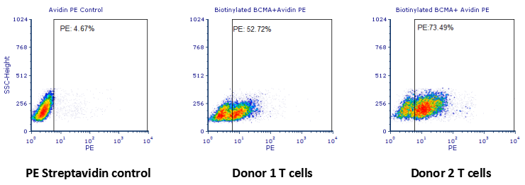 Human T cells were transfected with anti-BCMA CAR and cultured for 3 days. Three days post-transfection, 1x10⁶ cells were first incubated with 50ul biotinylated human BCMA protein (Cat. No. BC7-H82F0, 8ug/ml ), washed and then stained with PE Streptavidin and analyzed by flow cytometry.
 Data are kindly provided by PREGENE Biopharma
Case No.2 Evaluation of anti-CD19 CAR expression using FITC-labeled CD19 protein
-
Reagents:FITC-labeled Human CD19 (20-291) Protein (ACROBiosystems, Cat. No. CD9-HF2H2).
-
Samples:Anti-CD19 CAR-293 cells.
-
Brief Protocol:
1.Culture anti-CD19 CAR-293 cells in DMEM medium with 10% FBS in the CO2 incubator (at 37°C, 5% CO2). 2.Harvest the cells and wash the cells once by wash buffer. 3.Count the cells number and the viability, aliquot up 2e5 live cells (Anti-CD19-scFv positive cell is 98%) into each tube. (Note: the cell viability must be ≥ 95%.) 4.Add 100 µl, 10 µg/ml of FITC-labeled Human CD19 (20-291) Protein (ACROBiosystems, Cat. No. CD9-HF2H2) or FITC-labeled Protein control into each tube, incubating at 4℃ for 1 hour. 5.Wash the cells 3 times by wash buffer and resuspend the cells in 200 µl PBS per sample. 6.Transfer the cells into flow tube and detect by Flow cytometry. 7.Analyze result using FACS Celesta software and FCS Express 6 Flow software. -
Results:The data showed that the expression level of anti-CD19 scFv on the surface of anti-CD19 CAR-293 cells was 95.37%.
-
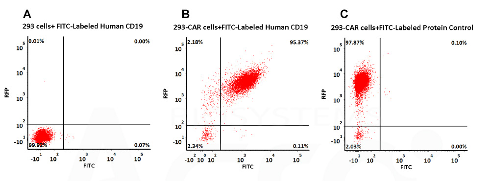 293 cells were transfected with anti-CD19-scFv and RFP tag. 2e5 of the cells were stained with B. FITC-Labeled Human CD19 (20-291) (Cat. No. CD9-HF2H2, 10 µg/ml) and C. FITC-labeled protein control. A. Non-transfected 293 cells and C. FITC-labeled protein control were used as negative control. RFP was used to evaluate CAR (anti-CD19-scFv) expression and FITC was used to evaluate the binding activity of FITC-labeled Human CD19 (20-291) (Cat. No. CD9-HF2H2).
Case No.3 Evaluation of anti-CD19 CAR expression using FITC-labeled anti-FMC63 scFv antibody
-
Reagents:FITC-Labeled Monoclonal Anti-FMC63 scFv Antibody, Mouse IgG1 (ACROBiosystems, Cat. No. FM3-FY45).
-
Samples:Anti-CD19 CAR-293 cells.
-
Brief Protocol:
1.Culture Anti-CD19 CAR-293 cells in DMEM medium with 10% FBS in the CO2 incubator (at 37 ℃, 5% CO2). 2.Harvest the cells and wash the cells once by FACS buffer. 3.Count the cells number and the viability, aliquot up 2e5 live cells into each tube. (Note: the cell viability must ≥ 95%.) 4.Dilute FITC-Labeled Monoclonal Anti-FMC63 scFv Antibody, Mouse IgG1 (ACROBiosystems, Cat. No. FM3-FY45) in FACS buffer to get the working solution just before the assay, and then add 100 μL of the working solution into the tube with cell pellet. Mix well and incubate at 4℃ for 60 minutes. 5.Wash the cells 3 times by FACS buffer and resuspend the cell pellet in 200 μL PBS per sample. 6.Transfer the cell suspension into flow tube and detect the cells by Flow cytometry. 7. Analyze the result data using FCS Express 7 Plus and GraphPad Prism 5 software. -
Results:The data showed that the expression level of anti-CD19 scFv on the surface of anti-CD19 CAR-293 cells was 100%.
-
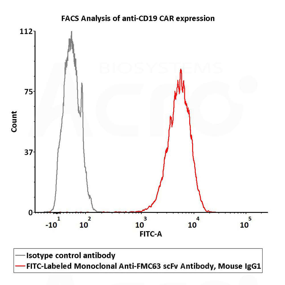 2e5 of Anti-CD19 CAR-293 cells were stained with 100 μL of 1:50 dilution (2 μL stock solution in 100 μL FACS buffer) FITC-Labeled Monoclonal Anti-FMC63 scFv Antibody, Mouse IgG1 (Cat. No. FM3-FY45) and isotype control respectively. FITC signal was used to evaluate the binding activity .
Case No.4 Evaluation of anti-MSLN CAR expression using PE-labeled MSLN protein
-
Reagents:PE-labeled Human Mesothelin / MSLN (296-580) Protein (ACROBiosystems, Cat. No. MSN-HP2H5).
-
Samples:Anti-MSLN CAR-293 cells
-
Brief Protocol:
1.Culture anti-MSLN CAR-293 cells in DMEM medium with 10% FBS in the CO2 incubator (at 37 ℃, 5% CO2). 2.Harvest the cells and wash the cells once by wash buffer. 3.Count the cells number and the viability, aliquot up 1e6 live cells into each tube. 4.Add 100 µl of diluted PE-labeled Human Mesothelin (296-580) Protein (Cat. No. MSN-HP2H5) (prepared in dilution buffer at 1:50 dilution) into each tube, incubating at 4℃ for 1 hour. 5.Wash the cells 3 times by wash buffer and resuspend the cells in 200 µl PBS per sample. 6.Transfer the cells into flow tube and detect by Flow cytometry. 7.Analyze result using FACS Celesta software and FCS Express 6 Flow software. -
Results:The data showed that the expression level of anti-MSLN scFv on the surface of anti-MSLN CAR-293 cells was 100 %.
-
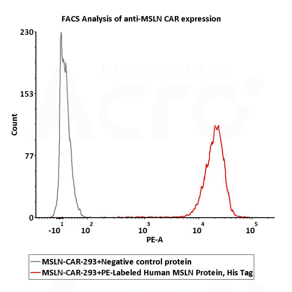 1e6 of the anti-MSLN CAR-293 cells were stained with 100 μL of 1:50 dilution (2 μL stock solution in 100 μL FACS buffer) of PE-Labeled Human Mesothelin (296-580) Protein, His Tag (Cat. No. MSN-HP2H5) and negative control protein respectively, PE signal was used to evaluate the binding activity.
Case No.5 Evaluation of the detection specificity of CD19, Fc Tag
-
Reagents:Human CD19 Protein, Fc Tag (ACROBiosystems, Cat. No. CD9-H5259);Human CD19 Protein ,Fc Tag (Company N);Human PD-L1 Protein, Fc Tag (ACROBiosystems, Cat. No. PD1-H5258), as negative control;FITC anti-human IgG Fc antibody (Biolegend, Cat. No. 409310).
-
Cells:R1013-C6 cells (Transfected 293 cells expressing the anti-CD19 [FCM63] scFv & RFP tag); Expi 293 cells; Jurkat E6.1 cells.
-
Brief Protocol:
1.Cells were stained for CAR expression using ACRO’s human CD19 protein, Fc Tag or Company N’s human CD19 protein, Fc Tag;2.After washing, secondary labeling was performed with FITC anti-human IgG Fc antibody;3.The cells were analyzed using NovoCyteTM flow cytometer and the data analyzed by FCS Express 6 Plus and GraphPad Prism 5 software. -
Results:The data shows that Company N’s human CD19-Fc fusion protein exhibits strong non-specific binding to 293 and Jurkat cells.
-
-
Binding specificity analysis of ACROBiosystems hCD19(C-Fc tag) protein
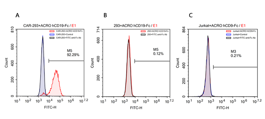 FACS analysis of human CD19 protein, Fc Tag (ACROBiosystems, Cat. No. CD9-H5259) binding to A. R1013-C6 cells, B. Expi 293 cells, C. Jurkat E6.1 cells. Cells were first stained with human CD19 protein, Fc Tag (ACROBiosystems, Cat. No. CD9-H5259) followed by FITC anti-human IgG Fc antibody, and then analyzed using NovoCyteTM Flow Cytometer. The data were analyzed with FCS Express 6Plus and GraphPad Prism 5 software.
Binding specificity analysis of Company N hCD19(C-Fc tag) protein
 FACS analysis of human CD19 protein, Fc Tag (Company N) binding to A. R1013-C6 cells, B. Expi 293 cells, C. Jurkat E6.1 cells. Cells were first stained with human CD19 protein, Fc Tag (Company N) followed by FITC anti-human IgG Fc antibody, and then analyzed using NovoCyteTM Flow Cytometer. The data were analyzed with FCS Express 6Plus and GraphPad Prism 5 software.
Reference
Related Articles
 Popular ArticlesRelated RecommendationsPopular Events
 Biomarkers: Enabling Precision Assessment of Cardio-Kidney-Metabolic (CKM) Syndrome2026-07-24Page Views:32
 Payload-Specific ELISA Kits for Intact ADC Quantification in Preclinical PK Studies2026-07-22Page Views:71
 Expanding the Potential of Hematopoietic Stem Cells (HSCs): Emerging Strategies for HSCs Expansion and Clinical Manufacturing2026-07-22Page Views:50
 Advances and Challenges of BCMA×CD3 Bispecific Antibodies in Multiple Myeloma2026-07-21Page Views:129
 E. coli Host Cell DNA Testing: Independent Eurofins Validation Demonstrates Reliable Residual DNA Quantification2026-07-20Page Views:109


![[Free Sample] Universal Fluorescent Anti-VHH Antibody — Precise Detection of VHH-Based CAR Expression](https://console.acrobiosystems.com/uploads/demo/2026/06/02/Anti-VHHAntibodySmallbanner.jpg)
 Related Products